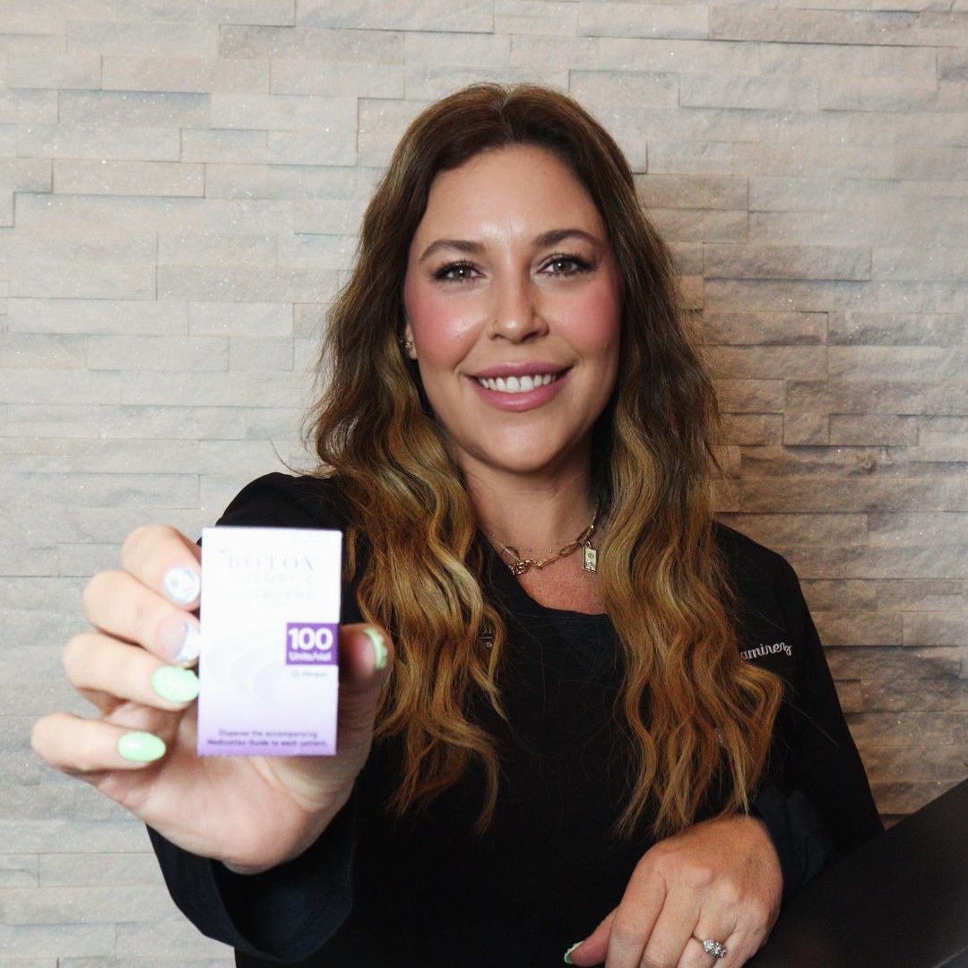
By Heather Ramirez, RN- Injector

Discover the hidden truths behind those viral MRI images and what they mean for your beauty routine
Yes, the viral MRI scan image showing dermal fillers has been making rounds on social media. It highlights how dermal fillers, which are commonly used in cosmetic procedures to enhance facial features, can be visualized in MRI scans. The image often sparks discussions about the visibility and longevity of these fillers in the body, as well as potential implications for those undergoing such cosmetic procedures.

So let’s get down to the information behind the topic.
When dermal fillers are visualized on MRI, their appearance can vary based on the type of filler used, the injection technique, and the timing post-injection. Over time, some fillers may appear more prominent on MRI due to changes in the filler material, water absorption, or surrounding tissue reactions.
Factors Affecting MRI Appearance:
1. Type of Filler: Different fillers (e.g., hyaluronic acid, calcium hydroxyapatite, or poly-L-lactic acid) have varying MRI characteristics. For example, hyaluronic acid fillers may appear hyperintense on T2-weighted images due to their water content.
2. Time Since Injection: Fillers may initially appear as distinct, well-defined areas on MRI. However, over time, the body’s reaction to the filler (such as encapsulation or fibrosis) might change its appearance. Some fillers might absorb water and swell, potentially doubling in volume, which could be seen as an increase in size on imaging.
3. Imaging Technique: Different MRI sequences (e.g., T1, T2, or STIR) might show fillers with varying intensities. The apparent volume on MRI can sometimes be misleading due to these differences in imaging sequences.
Doubling in Milliliters:
If a dermal filler appears to have doubled in volume on MRI (measured in milliliters), it could be due to:
– Hydration of the filler: Some fillers, particularly those based on hyaluronic acid, can absorb water over time, leading to an increase in their apparent volume.
– Inflammation or edema: Post-injection reactions might cause surrounding tissues to swell, which could be interpreted as an increase in the filler volume.
– Artifact or imaging differences: Variations in MRI technique or sequences might result in different measurements.
It’s important to interpret these findings in the context of the clinical scenario, including the type of filler used and the time elapsed since the injection.
In conclusion, choosing a trained and experienced provider for dermal filler injections is crucial. A qualified professional will not only have the expertise to administer the injections safely but will also select the appropriate products based on your specific needs, skin type, and desired outcome. This helps to minimize risks and achieve natural-looking, satisfying results.

